
[ad_1]
In the following sections, the results are discussed exclusively in terms of surface morphology and surface height variations as measured by laser profilometry. Any reference to mechanical, structural, chemical, or conservation-related phenomena is strictly limited to their observable surface expression and is intended solely to provide a morphological framework that may support and guide further investigations performed with complementary diagnostic techniques. These include subsurface imaging methods such as X-ray radiography (XR) or infrared reflectography (IRR), as well as spectroscopic approaches for chemical characterization, including X-ray fluorescence (XRF), Raman spectroscopy (RS), and Fourier Transform Infrared (FTIR) spectroscopy.
Supports
Figure 3 shows the 3D models of the four artworks, where surface topography is represented using a rainbow color scale ranging from blue (lowest points, set at 0.0 mm) to red (highest points, varying according to the sample). The macro-scale surface topography shown in Fig. 3 provides an overview of the surface height distribution across the full extent of the investigated artworks. These maps allow the visualization of large-scale surface height variations and spatial patterns that are primarily related to the geometry of the support and to surface features developed during the execution of the artwork. At this scale, the 3D reconstructions facilitate the identification of broad surface characteristics, such as height gradients, surface discontinuities, and preferentially oriented linear features, offering a comprehensive morphological context for the more detailed observations discussed in the following sections. Figure 3a presents the three-dimensional surface map of the oil painting on the wood panel. The 3D reconstruction clearly reveals the presence of the four horizontal wooden boards composing the support. The zero-reference level was assigned to the lowest point across the scanned area, located near the central joint where two boards were glued together. Additional minima were detected at the other junctions between boards. Laser profilometry also highlighted several natural characteristics of the wood structure. The maximum height variation of ~14.5 mm is attributable to the warping of the panel. These data are particularly valuable for monitoring shape or geometry changes of the wood over time and for assessing how the crossbars on the reverse side constrain or compensate for mechanical deformation. The analysis indicates that the containment effect is relatively effective in the upper region of the panel, but less so at the center. This suggests that mechanical deformations could be responsible for the formation of preferentially oriented linear surface features and, in some areas, visible cracks (Supplementary Fig. S1a). In solid mechanics, linear features observed on material surfaces are often discussed in relation to internal stress distributions. However, in the present study, such features are reported exclusively as surface height discontinuities measured by profilometry, without inferring internal stress states or mechanical forces, which should be measured through dedicated analytical techniques. An overview of the entire artwork is shown in Supplementary Fig. S1b, where the 2D color map is superimposed on the visible light image of the panel painting. As discussed, the crossbars on the reverse side help to limit changes in geometry of the support, particularly in the upper portion of the panel, while this effect is less evident in the central boards. In the image, the crossbars on the reverse are marked in black, whereas the linear surface features probably caused by the joining of the four boards and the cracks in the support are traced in white. These elements clearly distinguish areas under mechanical stress and provide valuable information about the structural integrity of the painting. Repeated profilometric measurements under varying environmental conditions and at different times would enable tracking of these linear surface irregularities and a more precise correlation between conservation state and environmental factors, ultimately creating a historical series of surface maps that are easily comparable over time.

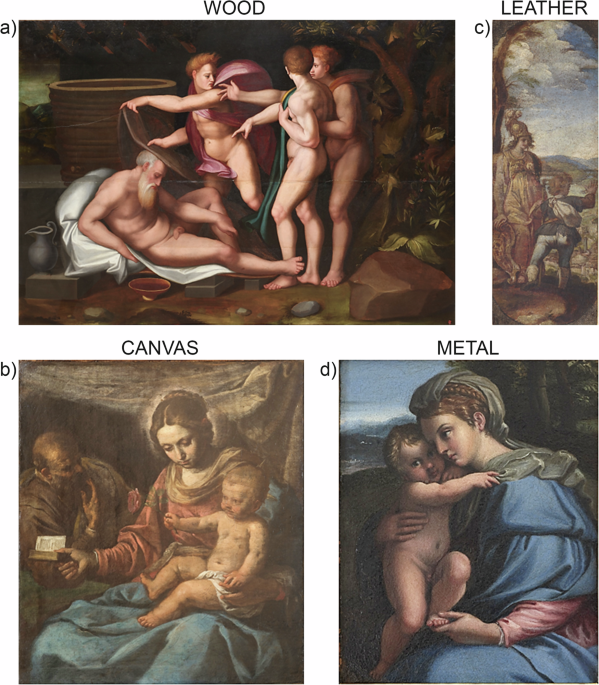
Orthogonal view of the 3D topographic images for the following artworks realized on different supports. a L’Ebbrezza di Noè (170 × 120 cm, oil on wood panel, 16th century); b La sacra famiglia (60 \(\times\) 50 cm, oil on canvas, 17th century); (c) Minerva indica a Taddeo la città di Roma (16 \(\times\) 45 cm, oil on leather, 16th century); (d) Vergine col Bambino (17 \(\times\) 21 cm, oil on copper, 17th century).
Surface height variations in the support are even more evident for the oil painting on canvas, shown in Fig. 3b. In this case, macro laser profilometry revealed regular planar areas but also depressions in the yarn that are not visually perceptible or detectable by touch. The zero-reference value was recorded in the central area of the painting, whereas the highest values (6–7 mm) were measured near the frame. The canvas thus appears slightly concave, suggesting a loss of tension toward the center. This change in the geometric property of the support may result from several factors, including natural deterioration of the canvas leading to fiber relaxation, improper stretching during mounting, or fluctuations in humidity and temperature. The difference in curvature between the perimeter, anchored to the frame, and the central area is a well-known phenomenon; however, its quantitative visualization, particularly in a relatively small and apparently rigid canvas, represents highly valuable information.
No significant alterations were detected in the oil painting executed on the leather support (Fig. 3c). This is likely due to the fact that the leather is mounted on a wooden frame, which provides greater mechanical stability to the artwork. At first glance, the surface topography appears fairly regular, with a maximum height variation of about 5.3 mm occurring at the junction between the two leather pieces. This is observable to the naked eye only on the reverse side of the artwork (Supplementary Fig. S2a). Overall, the sample exhibits relatively low surface roughness (data not shown), which can be attributed to the presence of a metallic silver leaf applied to the leather to create a smooth surface. Nevertheless, the surface shows at the micrometer scale, the characteristic wrinkles probably induced by the natural chemical modification process of the leather, which mainly tends to dehydrate over time32; such a process imparts a slightly rough appearance, perceptible even beneath the layers of glue, silver, paint, and varnish. Additional valuable information was also obtained from the scanning of the verso. As previously noted, the reverse side of the leather support clearly reveals the junction between the two leather sections, likely created through stitching or gluing, and the 3D topographic image highlights this fabrication process (Supplementary Fig. S2b). Finally, a detail of historical significance was also documented: a seal bearing the emblem of the Santacroce family, the former owners of the artwork (Supplementary Fig. S2c, d). The digital acquisition of such features is of particular interest to art historians, as it provides a higher level of detail and reproducibility compared to conventional visible-light photography.
Figure 3d presents the topographic map of the oil painting executed on a metallic support. Unlike the previous cases, the reconstructed image clearly reveals, even at the macroscopic scale, the contours of the depicted figure as slight surface reliefs. The surface topography appears highly homogeneous, with a maximum height variation of only about 2.5 mm. This suggests that the metallic substrate used for the artwork was manufactured with a very smooth finish, causing the painted figures to emerge in relief relative to the background. Copper supports typically have a thickness ranging from 0.5 to 2.0 mm and are produced either by hammering or by rolling thin sheets of metal, later trimmed with specialized shears. Hammered sheets generally show a more irregular texture with pronounced protrusions, whereas rolled sheets exhibit a smoother and more uniform surface, as they are compressed between two rollers33,34. The 3D model shown in Supplementary Fig. S3 suggests that the plate used in this case could be formed by rolling, and it is remarkable how the profilometric data preserve the “material memory” of this technological phase in the artwork’s production.
Paint layers
Macro laser profilometry provides quantitative information on the surface morphology of paint layers, allowing the documentation of surface features related to painting execution and surface condition. In this context, the technique is used exclusively as a spatially resolved surface reference, which can support and guide the targeted application of complementary imaging and analytical methods when information on material composition or subsurface structure is required. Figure 4 presents surface profilometry maps magnified to highlight surface features at the micrometer scale. Compared to the macro-scale observations in Fig. 3, the magnification of surface 3D maps enables a more detailed visualization of fine surface features of the paint layers, including micro-scale height variations, shallow surface discontinuities like micro-cracks, and features related to painting execution, such as brushstroke morphology. In some cases, image-processing filters were applied to the height maps (e.g., Figs. 4b and 5) to enhance edge contrast and facilitate the visualization of subtle surface details. The visible light and height images of a detail from the panel painting are shown in Figs. 4a and b, respectively. Unlike the rainbow-scale topographic maps, the height images are derived from the calculation of intensity differences between adjacent pixels, producing a pseudo-relief effect that effectively enhances micro-details such as incisions, scratches, and brushstrokes. In this case, the height map reveals incisions in the preparatory layer (indicated by yellow lines), appearing randomly distributed across the surface. These marks may be accidental or intentionally made to improve the adhesion of the paint layers to the ground layer. The application of the emboss filter further accentuates the surface morphology of the brushstrokes, providing valuable insights into the artist’s working process. Based on the observed sequence, the preparatory layer was first applied and incised, followed by the outlining of the drawing, the filling of the background, and the definition of the figures’ contours. The composition was then refined with glazes over the flesh tones to enhance modeling and detail, and finally, highlights were added as the concluding step.

Visible light (a) and height (b) image of a detail from painting on wood panel; 3D topographic images of: central part of the canvas (c), detail of the upper side of the canvas (left and middle) and line profile (right), central (d) and lower (e) part of the leather painting.

a Front side, height image superimposed to the visible light image; (b) detail in visible light image; (c) detail of the height image elaborated through emboss filter; (d) reverse side, height image; e detail of the reverse side of the painting.
Figure 4c presents the 3D rainbow-scale topographic maps of the central portion of the canvas (left) and a detail of its upper area (middle). A clear difference can be observed between the left and right sides, with the right side appearing thicker, likely due to the presence of an underlying composition covered by a second paint layer. This thickness variation is also evident in the corresponding line profile (Fig. 4c, right), which displays a distinct step associated with the repainting (highlighted by a red circle). Furthermore, a noticeable contrast in surface morphology is observed: on the left side, the canvas weave remains visible beneath the paint layers, whereas on the right side, deep cracks are present (Fig. 4c, center). In this case, the step is particularly pronounced and even perceptible to the touch. However, the quantitative measurements provided by profilometry allow for a more detailed interpretation, potentially revealing subtle thickness variations that might not be visually detectable and could be related to hidden voids rather than overlapping paint layers.
For the painting executed on a leather support, the analysis focused on the central and lower portions of the artwork, as shown in Fig. 4d and e, respectively. As previously mentioned, the surface topography image reveals the junction between two pieces of leather. Interestingly, this joint is not visible to the naked eye but can be observed on the reverse side of the painting. The 3D map clearly highlights this junction, which, despite being covered by paint layers, is easily detected through macro laser profilometry. This observation also provides insight into the fabrication process, which typically involves abrading the leather surfaces and then joining them using animal glue. Moreover, the wrinkling of the support also extends to the pictorial surface, where the loss of underlying material in some areas has led to localized paint losses (circled detail in Fig. 4d). In the lower portion of the painting (Fig. 4e), the scanning process revealed decorative motifs impressed in the corners, likely created using metallic punches, a common technique in leather decoration35,36. In this case, the punches appear to be either square, composed of three circular elements per side, or rectangular, featuring two columns of three circles. The stamping was evidently carried out prior to the application of paint, as some of the geometric motifs are partially covered by the painting layers.
The results obtained for the painting on the copper support are presented in Fig. 5. The height image in Fig. 5a shows that the different color fields correspond to varying states of conservation, possibly related to differences in the pigment-to-binder ratio. In fact, it is well known in the literature that the aging of the oil binder can induce chemical and physical modifications in the paint layer, often associated with shrinkage during drying and the subsequent formation of cracks. The craquelure pattern is more pronounced in darker areas, where pigments absorb a larger amount of binder, and less evident in lighter regions such as flesh tones, which generally exhibit faster drying properties. Furthermore, when adhesion between the paint layer and the copper substrate is poor, raised areas may appear on the painted surface, indicating partial detachment. In more severe cases, complete detachment can occur, leading to flaking or lifting of the paint film32,33. Figure 5b, c highlights such detached regions (indicated by red arrows), which are clearly visible in the height image but not discernible in the visible light image due to overlying retouches. Figure 5d shows the height map of the verso of the painting, where numerous incisions made on the copper plate are traced in yellow. The detail in Fig. 5e illustrates both exposed and covered incisions, providing valuable information about the preparation of the metallic support. Copper plates were traditionally roughened with sharp tools or abrasive paper to improve paint adhesion. Additional details from the reverse side are shown in Supplementary Fig. S3. Supplementary Fig. S3b reveals slight deformations of the plate caused by mechanical pressing during manufacture, while Supplementary Fig. S3c displays a fingerprint, presumably left unintentionally by the artist. This imprint not only documents the creative process but also represents a tangible trace of the artist’s physical presence, adding an intimate and historically significant dimension to the artwork.
In conclusion, this study demonstrates the effectiveness of macro laser profilometry as a quantitative technique for documenting surface morphology and surface height variations of artworks realized on different supports. This approach enables the acquisition of a wide range of detailed information using a single instrument, thereby eliminating the need to switch between tools for different spatial scales and ensuring greater consistency in the collected data. The application of laser profilometry to painted surfaces offers several advantages, as summarized in Table 2. The technique is entirely contactless, non-destructive, and non-invasive. Its portability allows for in-situ investigations, which is particularly valuable for artworks that cannot be moved. Furthermore, the high-speed acquisition capability and automation of the scanning process significantly reduce measurement time while enhancing reproducibility. The resulting 3D surface maps allow detailed documentation of surface features related to the nature of the support (wood, canvas, leather, or metal) and the execution of the pictorial layers, providing a robust and quantitative reference for surface documentation. The method also enables long-term monitoring of changes in the macro-geometry of the support and the micro-geometry of the pictorial film over time. Despite these advantages, the technique also presents certain limitations. The profilometer is sensitive to vibrations, and reflections from metallic or highly varnished surfaces can introduce spurious data points, affecting measurement accuracy. For large surfaces, multiple scans are required, which increases acquisition and processing time and demands substantial computational resources for data elaboration and analysis. This experimental study also demonstrates the benefits of integrating automated acquisition and data processing through Python-based tools, which streamlined the workflow and improved repeatability. The resulting 3D visualizations facilitate data interpretation for conservators and art historians. Moreover, the method supports rapid and quantitative comparisons of artworks under different environmental conditions, providing valuable information for diagnostic assessment and preventive conservation.
[ad_2]
Source link